BioPhysics Approach: Rheology & Microscopy
Key Skills Acquired
Rheology tests for biological fluid, including plotting and analyzing data.
Various microscopy techniques, such as confocal and spinning disk microscopy.
Effective communication with team members from diverse fields.
Live-cell imaging, and improvising methods for specific goals.
Strong interdisciplinary collaboration skills.
Rheology
During my Ph.D. training, I studied how Giardia, a motile protozoan intestinal parasite, influences and also be influenced by the host environment during infection. My biophysical approach involved studying how Giardia cells respond to the mechanical stress in the gastrointestinal tract. Specifically, I used rheology to test the mechanical properties of different fluids, such as viscosity and elasticity, to mimic the in vivo mechanical stress created by mucus in a laboratory setting.
This experience was immensely rewarding. I not only learned the specific skills of how to determine the mechanical properties of materials but also how physicists approach scientific questions. Through extensive collaborations, I gained valuable insights into explaining biological research to physicists or audiences in different fields and vice versa. This interdisciplinary experience has instilled in me a habit of always considering the physical aspects of biological research, which is invaluable for my future investigations.
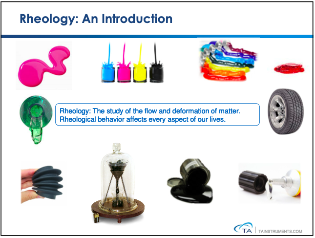
Figure 1: Rheology: Study of the flow and deformation of materials under force.
Microscopy
Another critical aspect of my research was microscopy, which I used to capture various properties of Giardia cells, such as attachment and motility behaviors. I gained experience with different microscopic techniques to achieve specific research aims. Below is an example of a live Giardia trophozoite attaching in culture, captured using a confocal microscope to collect motility details under treatment conditions.
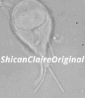
Figure 2: Confocal image of a _Giardia_ trophozoite in culture.
Here is another example of a live Giardia image obtained through a spinning disk microscope. This technique allowed me to capture the detailed structures of the 12-15µm long, 2µm thick cell, during the dynamic beating motion of its flagella.
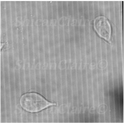
Figure 3: Live _Giardia_ trophozoite captured with a spinning disk confocal microscope.
Determining Fluid’s Rheological Properties
A significant part of my research involved testing and analyzing the viscosity, elasticity, and shear thinning or thickening properties of fluids using a rheometer. Below are some examples:
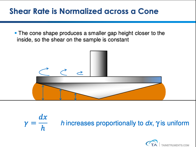
Figure 4: A cone and plate geometry rheometer used to determine fluid viscous and elastic moduli by increasing shear frequency or deformation rate.
We determined a material’s shear thinning or thickening properties by observing how its viscoelasticity changes with increased shear rate.
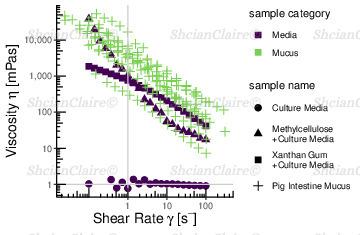
Figure 5: Flow curves showing shear thinning properties with various profiles for the tested fluids.
Additionally, we assess a material’s viscous modulus (loss modulus, G”) and elastic modulus (storage modulus, G’) by applying shear with increased frequencies. When the storage modulus exceeds the loss modulus, it indicates that the fluid’s elastic properties dominate at that frequency.
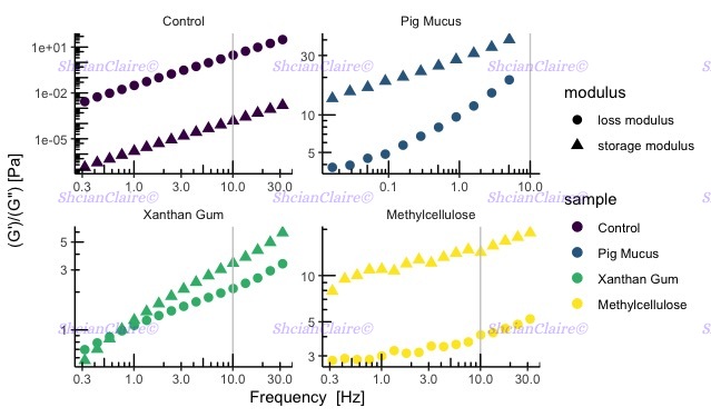
Figure 6: Examples of G’ and G” for various fluids.
Plot examples are original. The publication is currently under review.